|

|
No. 95 June 2014 |
|
||||
| |
Innovation and Development
Brand New Theory Realizes Sharp X-ray Visualization of Vascular Network
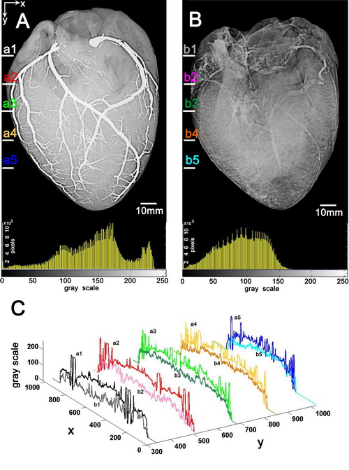
A joint team from the Technical Institute of Physics and Chemistry, CAS and Tsinghua University led by Prof. Liu Jing has proposed for the first time a room temperature liquid metal angiography to achieve mega contrast X-ray visualization of vascular network. The relevant work has been published in IEEE Transactions on Biomedical Engineering (Wang Q, et al., Liquid Metal Angiography for Mega Contrast X-ray Visualization of Vascular Network in Reconstructing In-vitro Organ Anatomy, 2014). According to the research, the liquid metals, such as gallium can be kept in liquid state at room temperature and infused into the vessels without destroying the tissues. While endowed with a high density, the liquid metal-filled vessels would generate a magnificent contrast with the surrounding tissues under the CT scan or X-ray photography. The method is even feasible to map tiny capillaries owing to the fluidity and compliance of the liquid metal. It was found in the experiments that the reconstructed vessels of in vitro pig hearts and kidneys infused with liquid metal gallium were extremely clear in the images. Generally speaking, the image contrast in the liquid metal angiograms was orders higher than that of the conventional iohexol enhanced images while the former still disclosed much richer vessel details. Further, the contrast effect in liquid metal angiograms would not gradually decay over time, which was in fact a weak point lying behind the traditional contrast agents. This work had ever aroused tremendous attentions throughout the world. In the last November 2013, when the authors posted their work to the web of arXiv.org, a series of world renowned scientific or technical websites such as Medium, Gizmodo and Slashdot etc. had ever made in-depth reports on the research by the story reads as “First Images of a Heart Injected with Liquid Metal”. Except for serving as a powerful angiographic tool for mapping the vasculature, the present image reconstruction principle can also be extended to more scientific or engineering areas where quickly reconstructing high resolution spatial channel networks is critical. On condition that the resolution of the imaging apparatus is high enough, the tiniest channel that can be distinguished by the current method could even reach nano meter scale.

copyright © 1998-2015
CAS Newsletter Editorial Board: 52, Sanlihe Road, Beijing 100864,
CHINA
Email: slmi@cashq.ac.cn