[Image: Cheng Zhen’s laboratory at CAS’s SIMM and Han Xiao’s laboratory at Rice University]
Near-infrared region two (1,000-1,700 nm, NIR-II) fluorescence imaging is a promising strategy for theragnostic and imaging-guided surgery. Compared to visible and near-infrared region one (600-900 nm) imaging, NIR-II imaging shows significant advantages, including less tissue autofluorescence background, deeper tissue penetration, and higher image resolution. In recent years, the investigation of NIR-II probes and their applications in the theragnosis of disease has experienced lots of attention and development. NIR-II dyes with large molecular frameworks still have limited applications for brain imaging since the blood-brain barrier (BBB) prevents most molecules from entering the central nervous system. Therefore, developing NIR-II fluorescence probes with BBB penetrability is desired for noninvasive brain imaging.
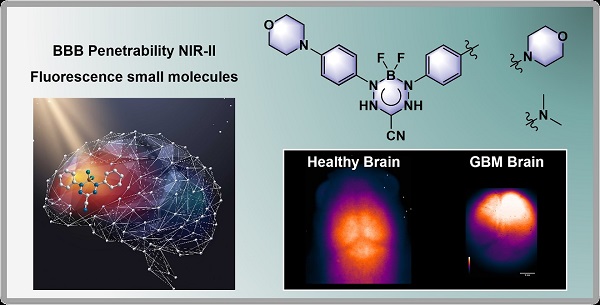
In a study published in Journal of the American Chemical Society (JACS) on December 2022, Cheng Zhen’s group from the Shanghai Institute of Materia Medica (SIMM) of the Chinese Academy of Sciences (CAS) and Han Xiao’s group from Rice University together developed two NIR-II dyes with BBB penetrability for noninvasive brain imaging and differentiating of normal brain tissue from glioblastoma tissue in vivo.
The series of boron difluoride (BF2) formazanate NIR-II dyes exhibited broad emission bands ranging from 800 to 1,400 nm, large Stokes shifts, strong fluorescence intensity beyond 1,000 nm and relatively high quantum yields. They also show attractive physiochemical parameters for BBB crossing, such as their small molecular weight (< 488 g mol-1) and appropriate lipophilicity (1.3 < log P < 3.2). To further evaluate the BBB permeability of BF2 formazanate dyes, an in vitro BBB model prepared by seeding bEnd.3 cells onto gelatin-coated upper chambers of the Transwell plates was used. BF1 and BF6 with morpholine and dimethylamine moieties exhibited significantly higher BBB permeabilities among the BF2 formazanate dyes tested.
To evaluate the use of the dyes for NIR-II imaging of cerebral tissue, researchers used tail vein injections to introduce BF1-BF8 dyes into athymic nude mice, followed by in vivo NIR-II fluorescence imaging. Significant BF1 and BF6 fluorescence signals were observed in brain tissue after injection, and sharp cerebral images with high signal to-noise ratios (SNRs) of 2.5-2.9 were obtained. The researchers observed the process of dye diffusion through the BBB, and both BF1 and BF6 showed rapid diffusion from vessels into brain tissue in 90 seconds.
Then BF1 and BF6 dyes were used for imaging the mouse model of GBM. By comparing the brain fluorescence images, researchers found that normal brain tissue exhibits a higher fluorescent intensity than GBM tissue. Most importantly, these results demonstrated that normal and brain tumor tissues were easily differentiated and delineated at the margins using in vivo and ex vivo NIR-II fluorescence imaging. Compared with indocyanine green (ICG), brain images of BF1- and BF6-treated mice established a significantly larger normal-to-glioma ratio (NGR), suggesting that BF1 and BF6 can better differentiate normal tissue from glioma than ICG. These results indicate that BF2 formazanate dyes are potential tools for differentiating GBM tissue during different growth times, thus facilitating clinical image-guided intervention.
We anticipate that this new type of small molecule will find potential applications in creating probes and drugs relevant to theranostics for brain pathologies.
For more information, please contact:
Diao Wentong
E-mail: diaowentong@simm.ac.cn
Shanghai Institute of Materia Medica,
Chinese Academy of Sciences
Source: Shanghai Institute of Materia Medica,
Chinese Academy of Sciences