Recently, an encouraging artificial intelligence (AI) technology for clinical application was announced by the teams at the Institute of Automation, Chinese Academy of Sciences (CAS), the Third Affiliated Hospital of Sun Yat-sen University, and the Chinese PLA General Hospital. Twelve hospitals in China got involved in this particular multicenter perspective study to answer an important question. Can AI achieve accurate diagnosis of the liver fibrosis stage in chronic hepatitis B patients? The quick answer is YES.
HBV infection is a serious problem in China, with more than one-third of the world’s HBV-infected people (approximately 93 million) residing in this country. Liver fibrosis is a progressive condition in chronic hepatitis B (CHB), and the accurate assessment of fibrosis is essential for prognosis, surveillance and management of patients with CHB. Liver biopsy is considered the reference standard for hepatic fibrosis staging. However, it is invasive, non-repeatable, and limited by sample errors, inter-observer variability, and various potential complications. Therefore, a non-invasive imaging diagnosis with excellent accuracy has been desired for a long time in clinical practice.
To solve this problem, a special squad was formed by AI scientists from CAS and doctors from ultrasonic departments in hospitals. About 400 CHB patients with 2,000 two-dimensional shear wave elastography (2D-SWE) images were collected from 12 hospitals all over China. Then, a specially designed computer program adopting the deep learning technology was applied to perform an automatic analysis of all images. This particular program can extract a large variety of features included in multiple hidden layers of these images, not visible to the human eye, and then conduct a sophisticated quantitative analysis to classify the liver fibrosis stage of every individual. Once the program was thoroughly refined, hundreds of randomized image data were used to verify its diagnostic accuracy, and the results were very exciting.

Illustration of the deep learning process for analyzing 2D-SWE images of CHB patients
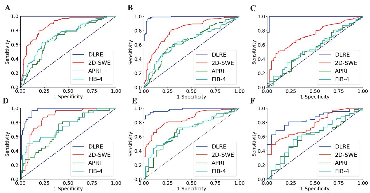
Compared with conventional ultrasonic elastography, the AI diagnosis offered a more than 15% increase in overall accuracy. For classifying cirrhosis and advanced fibrosis patients, it even provided diagnostic efficacy similar to the reference standard liver biopsy. Besides these results, many other unique advantages of using AI and ultrasound images for classifying liver fibrosis were also demonstrated in this study. But, maybe the most important finding is that this multi-disciplinary integration study convincingly proved the great potential of AI technology for helping clinical doctors in their daily routine practice, so that precision medicine can eventually become reality.
Dr. Wang Kun and his co-authors have published this work in GUT, 2018; 0: 1-13 (doi:10.1136/gutjnl-2018-316204, SCI IF: 16.658).
Source: Institute of Automation, CAS
For more information, please contact:
Dr. Wang Kun
E-mail: kun.wang@ia.ac.cn