CHINESE ACADEMY OF SCIENCES

-

- Structural insights into Niemann-Pick C1 (NPC1)-mediated cholesterol transfer and Ebola infection
- (NO.117 June 2016)
- Issue NO:Research Progress Updated:30-04-2020
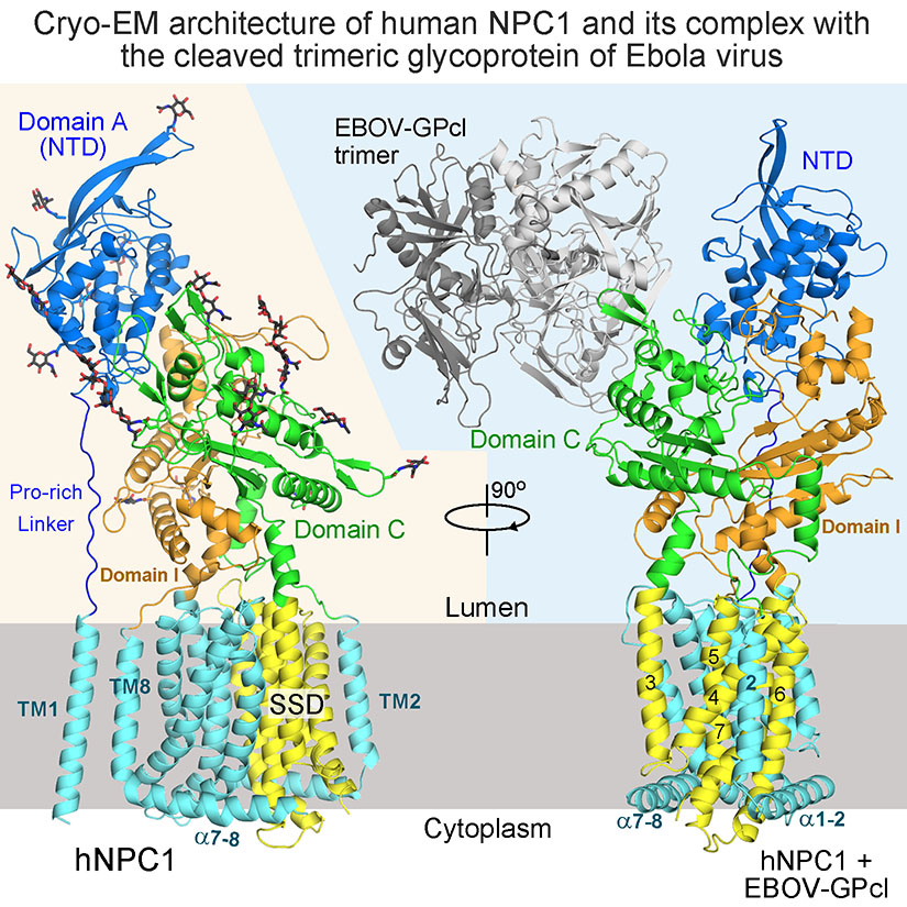
Niemann-Pick disease type C (NPC) is associated with mutations in NPC1 and NPC2, whose gene products are key players in the endosomal/lysosomal egress of low-density lipoprotein-derived cholesterol. NPC1 is also the intracellular receptor for Ebola virus (EBOV). Here, two research groups headed respectively by Prof. George F. Gao from the Institute of Microbiology, Chinese Academy of Sciences (CAS) and Prof. Yan Nieng from Tsinghua University present a 4.4 Å structure of full-length human NPC1 and a low-resolution reconstruction of NPC1 in complex with the cleaved glycoprotein (GPcl) of EBOV, both determined by single-particle electron cryomicroscopy. NPC1 contains 13 transmembrane segments (TMs) and three distinct lumenal domains A (also designated NTD), C, and I. TMs 2–13 exhibit a typical resistance-nodulation-cell division fold, among which TMs 3–7 constitute the sterol-sensing domain conserved in several proteins involved in cholesterol metabolism and signaling. A trimeric EBOV-GPcl binds to one NPC1 monomer through the domain C. Our structural and biochemical characterizations provide an important framework for understanding the mechanisms of NPC1-mediated intracellular cholesterol trafficking and Ebola virus infection.
The Niemann-Pick type C (NPC) lysosomal storage disease is characterized by the accumulation of cholesterol, sphingomyelin, and other lipids in endosomes and lysosomes. NPC patients may develop progressive neurodegeneration, hepatosplenomegaly, and premature death (Vanier, 2010 and Vanier and Millat, 2003). The disease is associated with NPC1 and NPC2 genes. NPC1 encodes a polytopic membrane protein that is located in the membranes of endosomes and lysosomes, and NPC2 is a small soluble protein in the lumen of endosomes and lysosomes or secreted from cells ( Carstea et al., 1997, Loftus et al., 1997 and Naureckiene et al., 2000). Genetic and biochemical characterizations revealed that NPC1 and NPC2 cooperate to export low density lipoprotein (LDL)-derived cholesterol from late endosomes and lysosomes to other cellular compartments ( Infante et al.,2008, Sleat et al., 2004 and Vanier, 2015).
The human NPC1 (hNPC1) consists of 1,278 amino acids (aa) and is comprised of 13 transmembrane helices (TM) and 3 distinct lumenal domains A, C, and I (Davies and Ioannou, 2000). The amino terminal (N) domain A is also designated NPC1(NTD). The prevailing model suggests that cholesterol derived from endocytosed LDL is extracted by NPC2, which may be recruited to NPC1 by domain C and deliver the bound cholesterol to NPC1(NTD) through a “hydrophobic handoff” mechanism (Deffieu and Pfeffer, 2011, Kwon et al., 2009 and Wang et al., 2010). The molecular basis for the ensuing intracellular trafficking of cholesterol remains largely elusive.
Sequence analysis suggested that NPC1 belongs to the resistance-nodulation-cell division (RND) superfamily (Davies et al., 2000, Scott and Ioannou, 2004 and Tseng et al., 1999), whose members, exemplified by the bacterial multidrug resistance pump AcrB and MexB, generally catalyze substrate export in change of H+ influx (Delmar et al., 2014, Venter et al., 2015 and Yamaguchi et al., 2015). NPC1 was reported to be a lipid exporter (Davies et al., 2000). TMs 3–7 of NPC1 were predicted to constitute the sterol-sensing domain (SSD), which is also identified in several other cholesterol metabolism or signaling-related membrane proteins, including the SREBP cleavage activating protein (SCAP) (Hua et al., 1996), the 3-hydroxy-3-methylglutaryl-CoA reductase (HMGCR) (Luskey and Stevens, 1985), NPC1-Like 1 protein (NPC1L1) (Altmann et al., 2004), and the Hedgehog signaling pathway proteins Patched and Dispatched (Burke et al., 1999, Hooper and Scott, 1989 and Nakano et al., 1989). The SSD acts as a regulatory domain in all these proteins (Doolman et al.,2004, Kuwabara and Labouesse, 2002, Malathi et al., 2004, Martín et al., 2001, Millard et al., 2005, Millat et al., 2001, Nohturfft et al., 1998, Strutt et al., 2001 and Yabe et al., 2002). Mutations in NPC1-SSD eliminated binding with a photoactivatable cholesterol analog and failed to crosslink with a small compound inhibitor for cholesterol egress (Lu et al., 2015 and Ohgami et al., 2004). The structure and functional mechanism of SSDs remain unclear.
In addition to cholesterol egress, NPC1 also serves as a critical component for cellular entry of Ebola virus (Carette et al., 2011 and Côté et al., 2011). The surface glycoprotein GP of the endocytotic viruses undergoes proteolytic cleavage by cathepsin (Chandran et al., 2005 and Schornberg et al., 2006). Binding of the cleaved GP (GPcl) to NPC1 through domain C is required for the subsequent release of the viral contents to the infected cells (Miller et al., 2012).
The crystal structures of individual NTD, domain C, NTD bound to sterols, and domain C in complex with GPcl of Ebola (EBOV-GPcl) were determined (Kwon et al., 2009, Wang et al.,2016 and Zhao et al., 2016). However, the structure of the transmembrane domain and how the distinct domains are organized remain to be elucidated. Here, we report the 4.4 Å structure of full-length (FL) hNPC1 and a 6.6 Å reconstruction of hNPC1 in complex with EBOV-GPcl, both determined using single particle electron cryomicroscopy (cryo-EM).
Their achievement was published on line on Cell on May 26, 2016. Authors include Gong Xin, QianHongwu, Shi Yi, George F. Gao,Zhou Qiang, Yan Nieng, et al.