CHINESE ACADEMY OF SCIENCES

-

- New technology for tracing cell proliferation developed and sources of adult hepatocytes discovered
- (NO.173 March 2021)
- Issue NO:Research Progress Updated:30-04-2021
On February 26, 2021 Beijing time, a study called “Proliferation tracing reveals regional hepatocyte generation in liver homeostasis and repair” was published in the journal Science. This study was conducted by Dr. Zhou Bin’s Lab at the Center for Excellence in Molecular Cell Science of the Shanghai Institute of Biochemistry and Cell Biology, Chinese Academy of Sciences. For the first time, scientists develop a new technology that enables long-term continuous monitoring of in vivo cell proliferation. By using this new technology, the researchers discovered the source of adult hepatocytes, shedding new light on liver regeneration research and clinical treatment of liver diseases.
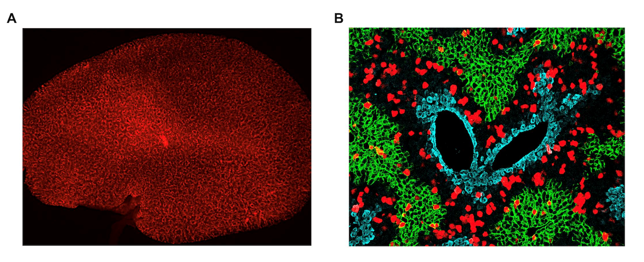
(A) Whole-mount fluorescence image of liver collected from hepatocyte-specific ProTracer mice; Hepatocyte proliferation events (tdTomato+ signal) occurred as a unique donut-shaped pattern. (B) Immunostaining for E-cadherin (E-CAD) (green), glutamine synthetase (GS) (blue), and tdTomato (red; ProTracer signal) on liver sections of hepatocyte-specific ProTracer mice; Hepatocyte proliferation events (tdTomato+) genetically recorded by ProTracer are highly enriched in the E-CAD?GS? midlobular zone during liver homeostasis. [IMAGE: CENTER FOR EXCELLENCE IN MOLECULAR CELL SCIENCE/SHANGHAI INSTITUTE OF BIOCHEMISTRY AND CELL BIOLOGY]
Cell proliferation is a fundamental biological process through which multiple-cell organisms undertake their development, tissue homeostasis, and regeneration. In many cases, disrupted proliferation is the pathogenic basis of many diseases. The ability to monitor cell proliferation has been essential for myriad studies in developmental biology, oncology, neuroscience, and regenerative medicine. Currently, two approaches are mainly used for analyzing cell proliferation in life science research: nucleoside-analog incorporation during DNA synthesis (such as BrdU and EdU), and tissue staining with cell proliferation markers (such as Ki67, pH-H3, etc.). However, the above two approaches have certain limitations: first, in terms of time, current approaches using marker staining can only detect cell proliferation at a single point in time rather than within a certain period of time. Like a camera, it can only provide a snapshot of cell proliferation signals. Thus, it’s difficult to apply the process to proliferation detection of certain types of cell with low proliferation capacity, such as cardiomyocytes and neurons; secondly, the proliferation signals of all cell types will be detected spatially and indiscriminately, which can easily introduce interference from the proliferation signals of cell types of little or no interest. Therefore, it’s important to develop a new technology to achieve continuous monitoring of cell proliferation in specific cell types.
Zhou Bin’s Lab has been committed to the development and application of new genetic lineage tracing technology for a long time. In this study, they took advantage of the widely used cell proliferation marker Ki67, and two orthogonal, site-specific recombinases systems, Cre-loxP and Dre-rox, to develop a genetic proliferation lineage tracing method — proliferation tracer (ProTracer). In this new technology, Dre-rox is used to prime the Cre-mediated cell proliferation tracing. Once activated, ProTracer can continuously record cell proliferation for months or even years, like a video recorder, which is very useful for detecting cells with low proliferation capacity and evaluating the dynamic changes of their proliferation. Moreover, ProTracer can be used to study proliferation of one specific cell lineage, avoiding interference from other types of proliferation signals and enabling observation of cell proliferation at the whole organ level. Furthermore, ProTracer permits non-invasive long-term monitoring of cell proliferation in live animals, providing researchers with a temporally seamless tool to investigate cell proliferation dynamics in the same animal over a period of hours, months or even longer. ProTracer could be widely used in multiple tissues and organs, providing powerful technical support for developmental biology, oncology, neuroscience and regenerative medicine and so on.
Researchers applied ProTracer to study the cell sources of adult hepatocytes during tissue homeostasis, repair, and regeneration, which is an important and controversial scientific issue. New hepatocytes are mainly derived from self-duplication in the adult stage. Hepatocytes are heterogenous due to their residence in different zones of the liver lobules, the functional units of the liver. The liver lobule is divided into three main distinct zones: the peri-portal zone surrounding the portal vein (marked by E-cadherin, E-CAD)-zone 1; the central zone nearest to the central vein (marked by glutamine synthetase, GS)-zone 3; and the mid zone-zone 2. It’s believed that the proliferation capacity of hepatocytes in these zones is quite different, but it remains unclear in which zone hepatocytes proliferate more preferentially. Currently, five different models of hepatocyte generation have been proposed: the streaming model (Hepatocytes flow from portal vein to central vein, Liver 1985); the pericentral hepatocytes model (Axin2+ hepatocytes, Nature 2015); the periportal hepatocytes model (Sox9+ hepatocytes, Cell 2015); the distributed hepatocytes model (Terthigh hepatocytes, Nature 2018); and the broad distribution model (modest proliferation of hepatocytes in all zones, Cell Stem Cell 2020). These previous studies mainly relied on a specific molecular marker to label a subpopulation of hepatocytes and trace their expansion, without analysis of the proliferation capacity of all hepatocyte populations.
Zhou Bin’s Lab developed a method to trace all hepatocyte populations rather than a subset of hepatocytes by applying ProTracer to label all the newborn hepatocytes, so that all the hepatocytes can be detected for their proliferation and the location where the new hepatocytes arise could be directly displayed. The researchers primed the ProTracer that can monitor global cell proliferation in adult mice by tamoxifen induction, and collected liver tissues at multiple time points; after that, immunostaining of GFP (new generated cells), GS (pericentral hepatocyte marker), E-CAD (periportal hepatocyte marker), and b-catenin (cell membrane marker) showed that newborn hepatocytes are mainly generated in the middle region of liver lobules.
In order to eliminate the interference of non-hepatocyte proliferation signals, researchers developed a hepatocyte-specific ProTracer by taking advantage of the previous ProTracer and the hepatocyte-specific expressed Albumin (Alb) gene promoter. The hepatocyte-specific ProTracer could discern the hepatocyte proliferation patterning from the whole organs. The team applied the hepatocyte specific ProTracer for detection of newborn hepatocytes. Whole-mount fluorescence imaging of livers showed that the newly generated tdTomato+ hepatocytes appeared as a unique donut-shaped pattern (Figure A). The results of immunostaining of liver sections confirmed this highly regional pattern, which indicates that the majority of hepatocyte proliferation activity had occurred in zone 2 (Figure B). Moreover, they developed a new version of ProTracer that can be extended for non-invasive monitoring of gradual hepatocyte proliferation recording events in live mice over the course of long-term studies; the first time cell proliferation detection in live animals has been possible.
To investigate new hepatocyte generation during liver regeneration, researchers examined multiple liver injury models, such as a partial hepatectomy (PHx) injury model, a carbon tetrachloride (CCl4) injury model, and a bile duct ligation (BDL) injury model, on ProTracer mice, and found that hepatocytes in zone 1 first responded to the PHx injury, and in zone 2 first responded to the CCl4 and BDL injury. They also systematically described the proliferation of other types of cells during liver regeneration, such as cholangiocytes, endothelial cells, fibroblasts and macrophages.
In summary, this work has developed a new technology — ProTracer for continuous monitoring and recording in vivo cell proliferation events during tissue homeostasis and regeneration. The study discovered the cell sources of adult hepatocytes during liver homeostasis and regeneration, shedding light on liver regeneration study and clinical treatment for liver disease. It provides powerful genetic tools for cell proliferation related research.
For more information, please contact:
Dr. & Prof. Zhou Bin
E-mail: zhoubin@sibcb.ac.cn
Center for Excellence in Molecular Cell Science,
Shanghai Institute of Biochemistry and Cell Biology,
Chinese Academy of Sciences
Source: Shanghai Institute of Biochemistry and Cell Biology,
Chinese Academy of Sciences